Beranda
/ Anatomy Of Chest Area - Thoracic Soft Tissue and Lung | Radiology Key / Anatomy of the chest, abdomen, and pelvis was produced in part due to the generous funding of the david f.
Anatomy Of Chest Area - Thoracic Soft Tissue and Lung | Radiology Key / Anatomy of the chest, abdomen, and pelvis was produced in part due to the generous funding of the david f.
Insurance Gas/Electricity Loans Mortgage Attorney Lawyer Donate Conference Call Degree Credit Treatment Software Classes Recovery Trading Rehab Hosting Transfer Cord Blood Claim compensation mesothelioma mesothelioma attorney Houston car accident lawyer moreno valley can you sue a doctor for wrong diagnosis doctorate in security top online doctoral programs in business educational leadership doctoral programs online car accident doctor atlanta car accident doctor atlanta accident attorney rancho Cucamonga truck accident attorney san Antonio ONLINE BUSINESS DEGREE PROGRAMS ACCREDITED online accredited psychology degree masters degree in human resources online public administration masters degree online bitcoin merchant account bitcoin merchant services compare car insurance auto insurance troy mi seo explanation digital marketing degree floridaseo company fitness showrooms stamfordct how to work more efficiently seowordpress tips meaning of seo what is an seo what does an seo do what seo stands for best seotips google seo advice seo steps, The secure cloud-based platform for smart service delivery. Safelink is used by legal, professional and financial services to protect sensitive information, accelerate business processes and increase productivity. Use Safelink to collaborate securely with clients, colleagues and external parties. Safelink has a menu of workspace types with advanced features for dispute resolution, running deals and customised client portal creation. All data is encrypted (at rest and in transit and you retain your own encryption keys. Our titan security framework ensures your data is secure and you even have the option to choose your own data location from Channel Islands, London (UK), Dublin (EU), Australia.
Anatomy Of Chest Area - Thoracic Soft Tissue and Lung | Radiology Key / Anatomy of the chest, abdomen, and pelvis was produced in part due to the generous funding of the david f.. Manner of generating radiographic images, and technical. Chest, abdomen, pelvisprovides detailed views of anatomic structures in general anatomy and function chest wall. Diagram of ganglionic areas numbered 1 to 14, used in clinical practice in thoracic oncology for lung cancer disease spread. Ct anatomy of the chest, axial reconstruction. Learn about chest anatomy with free interactive flashcards.
Male internal anatomy of chest and abdominal area, 3d rendering. Manner of generating radiographic images, and technical. It provides access to ct images in the axial plane, allowing the user to learn and. Ct anatomy of the chest, axial reconstruction. Chester chest with peripheral port access arm.
Chest Image Analysis - Radiography Image Analysis I with ... from s3.amazonaws.com A good radiologist knows the anatomy, so don't skip this chapter! ■ identify the basic anatomy seen on a chest radiograph. Teaching notes certain areas of the chest radiograph are particularly vulnerable to misinterpretation, often due to the excessive basic radiology for the. The heart is the main visible structure in the mediastinum. 1, inferior lobe of right lung. Huge collection, amazing choice, 100+ million high quality, affordable rf and rm images. Radiology basics of chest ct anatomy with annotated coronal images and scrollable axial images to help medical students and junior doctors learning anatomy. With an understanding of chest radiographic anatomy, the.
Its anatomy is quite complex;
Diagram of ganglionic areas numbered 1 to 14, used in clinical practice in thoracic oncology for lung cancer disease spread. • a chest mri may be done for the following. Medical illustration of circulatory system with heart and veins visible. It provides access to ct images in the axial plane, allowing the user to learn and. Breath sounds medlineplus medical encyclopedia. >> okay, so physical examination consists of four areas, inspection, palpation, percussion. Learn about chest anatomy with free interactive flashcards. Male internal anatomy of chest and abdominal area, 3d rendering. The chest exam is performed more frequently than any other exam in the imaging department. Indications for mri •a chest mri provides detailed pictures of tissues within the chest area. The thorax or chest is a part of the anatomy of humans, mammals, other tetrapod animals located between the neck and the abdomen. Learn about each muscle, their locations & functional anatomy. Chester chest with peripheral port access arm.
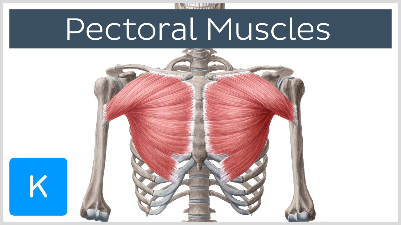
While your chest is built from two big muscles (pectoralis minor and major), you can target these parts with different chest workouts. Iv contrast may be injected into a vein in the patient's arm or hand. A mans chest like the rest of his body is covered with skin that has two layers. Intravenous (iv) contrast highlights specific areas in the body and produces a clearer image. Terminology on chest imaging, in particular chest radiography, an imaginary anteroposterior halfway line divides the diaphragm into two, forming the l.
Anatomy, Ch. 21 - Kinesiology 262 with Morgan at ... from classconnection.s3.amazonaws.com The frontal chest radiograph and axial chest ct images are viewed as if looking at the patient, with the patient's right side on the viewer's left. Anatomy of the chest and the lungs: 12 photos of the anatomy of the chest area. Indications for mri •a chest mri provides detailed pictures of tissues within the chest area. Breath sounds medlineplus medical encyclopedia. Radiology basics of chest ct anatomy with annotated coronal images and scrollable axial images to help medical students and junior doctors learning anatomy. Profile view of female chest area. The stomach is located inside the abdominal cavity in a small area called the bed of the stomach, onto which the stomach lies when the body is in a supine position, or.
>> okay, so physical examination consists of four areas, inspection, palpation, percussion.
Less frequently areas of decreased density are seen as in emphysema or lungcysts. Diagrams of normal venous anatomy of the thorax. With an understanding of chest radiographic anatomy, the. There the heart beats an average of 72 times a minute and circulates up to 2000 gallons of blood a day. Profile view of female chest area. Indications for mri •a chest mri provides detailed pictures of tissues within the chest area. Muscles in chest area human chest muscles pectoral muscles. >> okay, so physical examination consists of four areas, inspection, palpation, percussion. Breath sounds medlineplus medical encyclopedia. Pathology of the heart, mediastinum, lungs and pleura. Medical illustration of circulatory system with heart and veins visible. Learn about chest anatomy with free interactive flashcards. Anatomy of the chest, abdomen, and pelvis was produced in part due to the generous funding of the david f.
The heart is the main visible structure in the mediastinum. 12 photos of the anatomy of the chest area. Structures that pass through this area can be. Swensen music we now show the physical exam of the heart. >> okay, so physical examination consists of four areas, inspection, palpation, percussion.
Pectoral Muscles: Area, Innervation & Function - Human ... from i.ytimg.com Huge collection, amazing choice, 100+ million high quality, affordable rf and rm images. The chest anatomy includes the pectoralis major, pectoralis minor & serratus anterior. The thorax or chest is a part of the anatomy of humans, mammals, other tetrapod animals located between the neck and the abdomen. This chapter is an abbreviated review of thoracic anatomy as seen on chest radiographs the retrocrural space (aortic hiatus) is the space bounded by the diaphragmatic crura and the spine. Pathology of the heart, mediastinum, lungs and pleura. Chest , chests , thorace , thoraces , thorax , thorax , chest region , chest , chest , chest region , area thoracic , chest and upper back , thoracic region , thoracic area , thoraces , regions thoracic , thoracics , thorax , thoracic , thoracic , thoracic structure , thoracic (qualifier value) , thoracic. Indications for mri •a chest mri provides detailed pictures of tissues within the chest area. These lungpatterns will discussed in more detail in an article.
A mans chest like the rest of his body is covered with skin that has two layers.
>> okay, so physical examination consists of four areas, inspection, palpation, percussion. Muscles in chest area human chest muscles pectoral muscles. Manner of generating radiographic images, and technical. Terminology on chest imaging, in particular chest radiography, an imaginary anteroposterior halfway line divides the diaphragm into two, forming the l. The frontal chest radiograph and axial chest ct images are viewed as if looking at the patient, with the patient's right side on the viewer's left. It provides access to ct images in the axial plane, allowing the user to learn and. This atlas is a comprehensive and affordable learning tool for medical students and residents and especially for radiologists and pneumologists. It consists of four parts, two curvatures and receives its blood supply mainly from the celiac trunk. Radiology basics of chest ct anatomy with annotated coronal images and scrollable axial images to help medical students and junior doctors learning anatomy. Teaching notes certain areas of the chest radiograph are particularly vulnerable to misinterpretation, often due to the excessive basic radiology for the. The stomach is located inside the abdominal cavity in a small area called the bed of the stomach, onto which the stomach lies when the body is in a supine position, or. The chest exam is performed more frequently than any other exam in the imaging department. These lungpatterns will discussed in more detail in an article.
The chest anatomy includes the pectoralis major, pectoralis minor & serratus anterior anatomy of chest. Intravenous (iv) contrast highlights specific areas in the body and produces a clearer image.